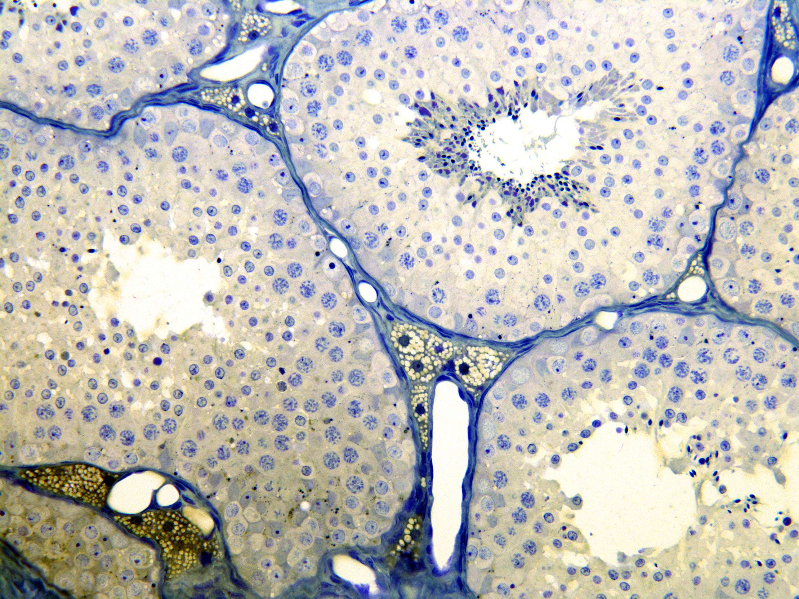
Testis from a cat (400X)
In the tubuli seminiferi in this section you see the different stages of maturation of the sperms (spermatogenesis) more detail. Cells in the first stage, which lie most peripherally, are the spermatogonia. The next stage is represented by the spermatocytes, and they are of similar size as the spermatogonia but have somewhat larger nuclei with coarse and darkly stained chromatin. The spermatides represent the next stage. They are much smaller and develop into the sperms, which you can see inside the lumen. Some Sertoli cells (mainly their nuclei) are visible in the periphery of the tubuli seminiferi. Their nuclei have an irregular shape, typically with the longest diameter perpendicular to the basal lamina of the tubules. The sperms (spermatozoa) have darkly stained, small and pointed heads that can be seen in or close to the lumen.
The interstitium is situated inbetween all the tubuli seminiferi. The intestitium houses lumps of Leydig cells. You can see that these cells contain some vacoules that have contained steroid hormone (testosterone). The steroids (lipids) have been removed during preparation in the central part of the section - therefore the vacouoles look light and empty. In more peripheral parts of the section (not seen in this photomicrograph) the lipid content is preserved, appearing as dark brown dots in the Leydig cells.


