
Testis (400X)
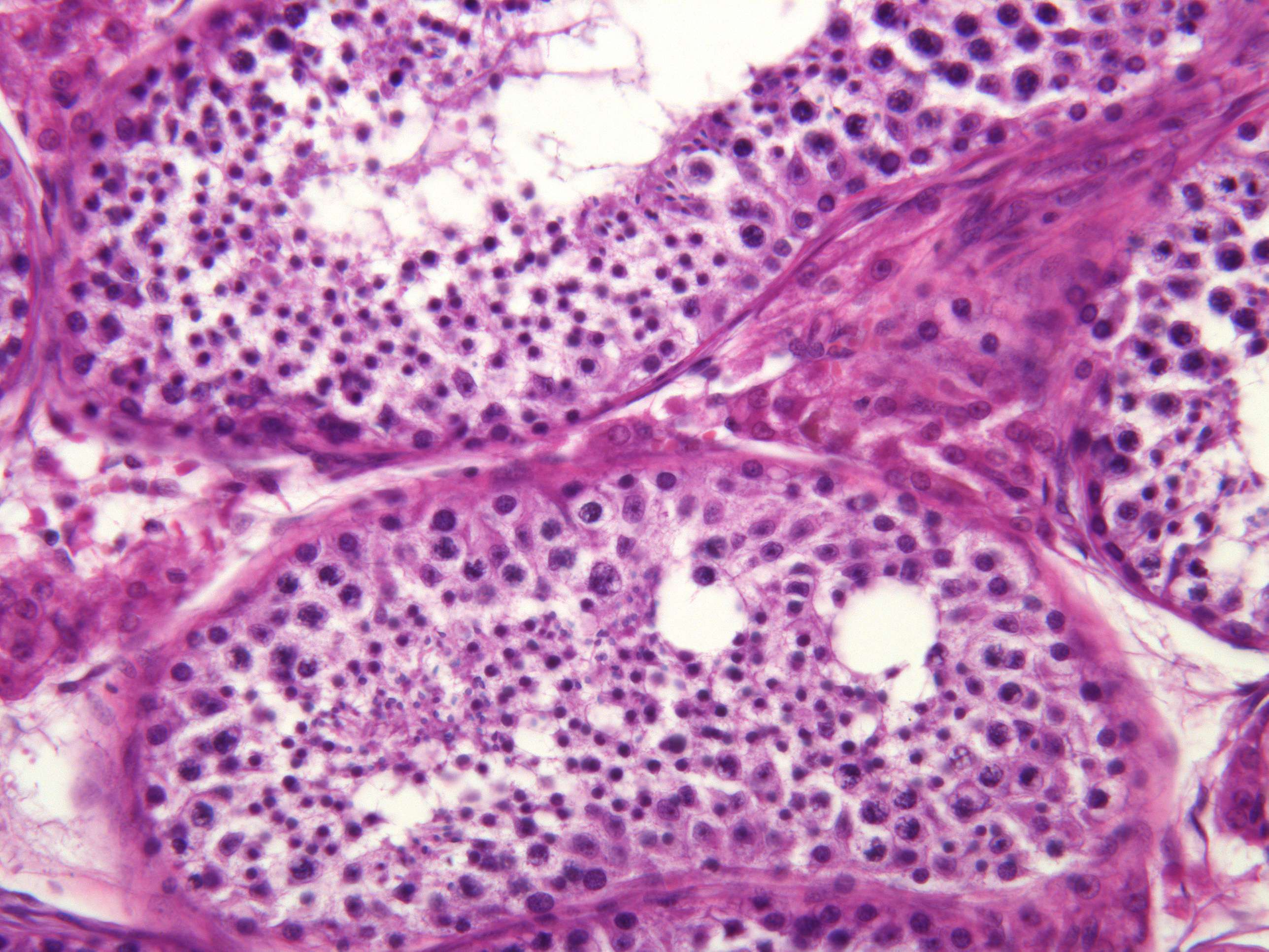
In this high power photomicrograph showing a seminiferous tubule and its surrounding interstitium, you can see many kinds of cells. From the basal membrane to the lumen you can find various stages of the spermatogenesis. The spermatogonia are located closest to the basal lamina. They are the "stem cells" and gives rise to all the other stages of developing sperms. Some spermatogonia enter the prophase of the first meiotic division, and remains at this stage for a long time. They move a little towards the lumen, and can recognized as primary spermatocytes (note their very coarse chromatin). The primary spermatocytes go through the first meiotic division, and each form two secondary spermatocytes. These go through the second meiotic division almost immediately and are therefore hard to find. The secondary spermatocytes give rise to spermatids, most of them found close to the lumen.
Among the cells in the various stages of the spermatogenesis, you can see Sertoli cells. This is a supporting cell; it forms the blood/testis barrier, supply nutrients to the developing sperms and phagocytizes cytoplasm that has been extruded from the spermatids.
Clumps of Leydig cells are seen in the interstitium between the tubuli seminiferi. Leydig cells produce the steroid hormone testosterone.