


Periodontal fibers and epithelial cell rests of Malassez (200X)
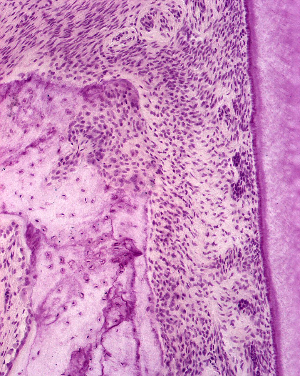
At the alveolar crest, you can see the orientation of the periodontal fibers. Note the numerous fibroblasts in the ligament. Close to the surface of the tooth, within the periodontal ligament, epithelial cell rests of Malassez can be seen. These epithelial cell rests can be distinguished from the surrounding tissue by their density and location and are named after Louis-Charles Malassez. The cells are remains of the Hertwig epithelial root sheath (HERS). It is believed that these cells, under special circumstances, contribute to the lining of different odontogenic cysts, i.e. the radicular cyst (periapical cyst).
The image is also well suited to study the alveolar bone and osteocytes. You can see how the periodontal fibers attach to the bone at the alveolar crest.