


Bud stage of tooth development in a human fetus (40X)
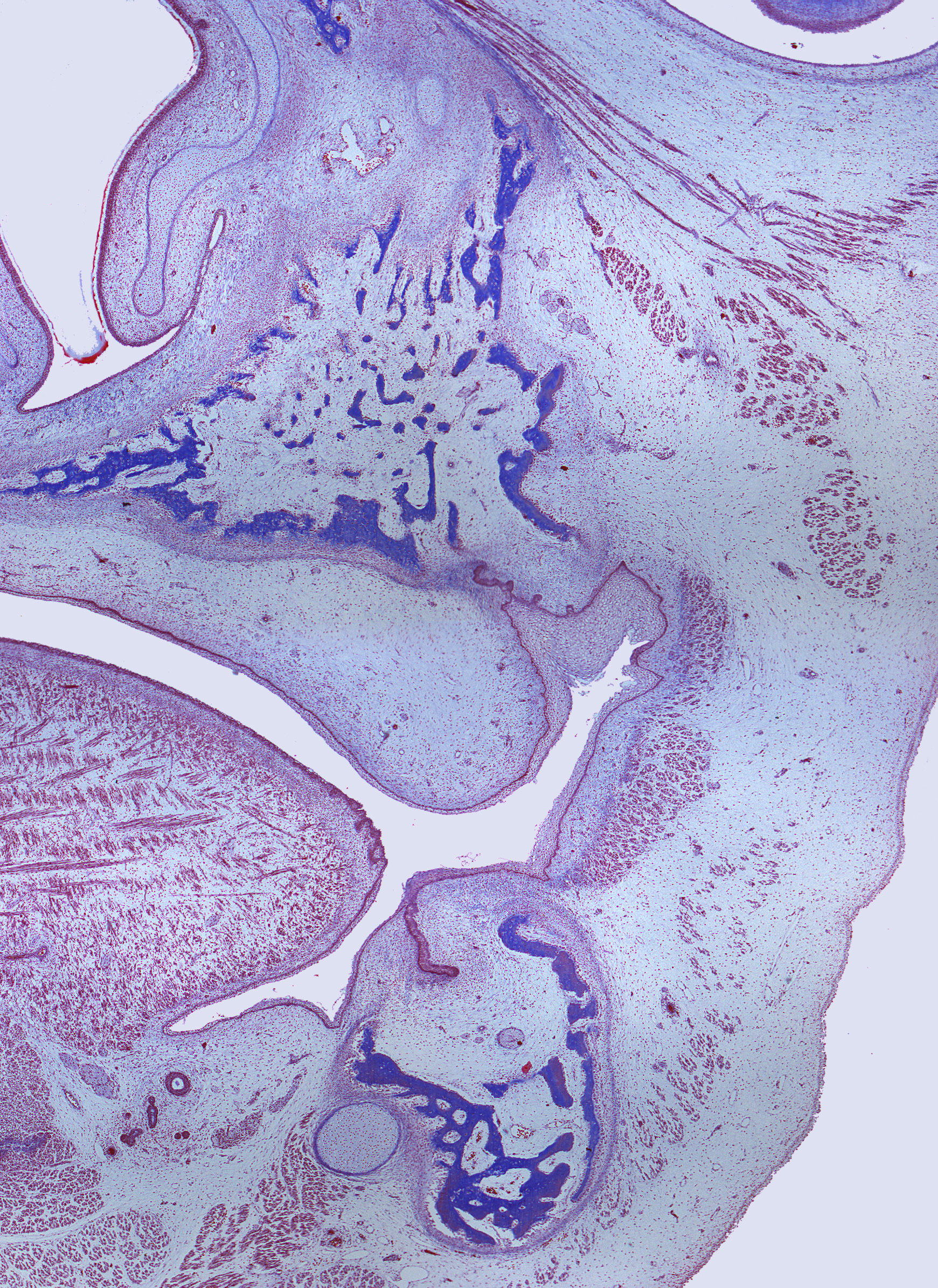
This is a frontal section of the head of a human fetus. Bone development has begun in both the maxilla and the mandible. Due to the stain used in this tissue sample, the bone appears blue.
Within the two quadrants visible here, there are two dental laminae, and surrounding these laminae, a condensation of mesenchyme is occurring. Between the spicules of bone in the mandible, you can see a cross-section of the alveolar nerve (n. alveolaris inferior in Latin). Meckel's cartilage is located medially to the mandibular bone. If you look closely, you can observe the downward growth of the salivary gland parenchyma and the developing muscle fibers of the tongue.
Press the 'magnify' button below the image to explore magnified areas of the section.